


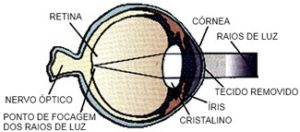
LASIK laser refractive surgery is the most advanced method for correcting refractive errors, particularly in the treatment of myopia, hyperopia and astigmatism. This surgical technique is a painless, fast and effective procedure that is performed with great safety.
The main goal of LASIK is to eliminate or reduce dependence on corrective lenses. This technique does not correct presbyopia or "tired eyesight", a natural condition that is directly associated with ageing and is based on the decreased ability to focus on close objects.
LASIK is the indicated method for those who suffer from myopia, hyperopia and/or astigmatism, are over 18 years of age and have healthy corneas. To perform this surgery, you should not have had a significant increase in the last 12 months, nor have any medical problems (specify which ones) that may prevent this operation.
The surgery consists of removing microscopic layers of corneal tissue, an equipment called Excimer Laser changes the shape of this component of the eye, allowing the rays of light to focus more accurately on the retina.
 In order to undergo this technique, you must get detailed information about your refractive defect by making a personal appointment and speaking to your doctor. If your refractive defect is in the LASIK correction range, some tests will be performed. If not, the doctor may recommend another refractive procedure in order to solve your problem.
In order to undergo this technique, you must get detailed information about your refractive defect by making a personal appointment and speaking to your doctor. If your refractive defect is in the LASIK correction range, some tests will be performed. If not, the doctor may recommend another refractive procedure in order to solve your problem.Before surgery:
The surgeon will evaluate if the patient meets all the conditions to perform the surgery, taking into account several aspects that can condition the results of this surgery, such as very dilated pupils, the eye being dry or previous ocular lesions.Procedure:
The patient lies in a reclining chair placed in an ambulatory surgery block. The eye to be operated on is anaesthetised with drops and then a small surgical instrument (blepharostat) is placed to hold the eyelids open during surgery. A suction ring is then placed over the cornea to fix it and prevent eye movement. At this time, the patient can feel the pressure of the blepharostat and vision may become duller. Next, the microkeratome - an automated microsurgery instrument - is attached to the suction ring. As the microkeratotome blade slides over the cornea the patient will hear a metallic sound and see poorly. When the microkeratotome stops working, a tissue sheet will be created which is folded back, becoming adherent at one end. (Fig. 1) Then, the laser is activated on the corneal thickness. At this point, the patient has to fixate a red light, while the vision is dull and hears a sound corresponding to the laser doing the ablation. (Fig. 2) Finally, the surgeon puts the flap that was folded back into place and it is fixed in position without the need for stitches (Fig. 3).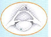
Fig.1
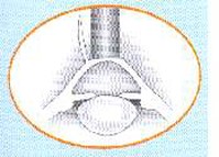
Fig.2
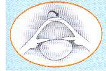
Fig.3
After surgery:
The patient will walk around with a bandaged eye for one day and will be able to resume their activity the next day. Vision will recover in the days or weeks following surgery and in some cases it may take several months to achieve the clarity of vision the patient had when wearing glasses or contact lenses.Like any other surgery, LASIK has risks and may have complications that, although very rare, should be carefully considered.
In some cases under- or over-corrections can occur. Fortunately, these can be corrected with spectacles, contact lenses or further surgery. There is still a small chance that the vision after surgery will not be as good as when wearing glasses or contact lenses.
Some people experience temporary discomfort, blurred vision, tearing, dry eye, dazzling, halos around lights, light sensitivity and fluctuating vision. These symptoms usually disappear within a month. In some cases they persist longer and may not disappear completely.
